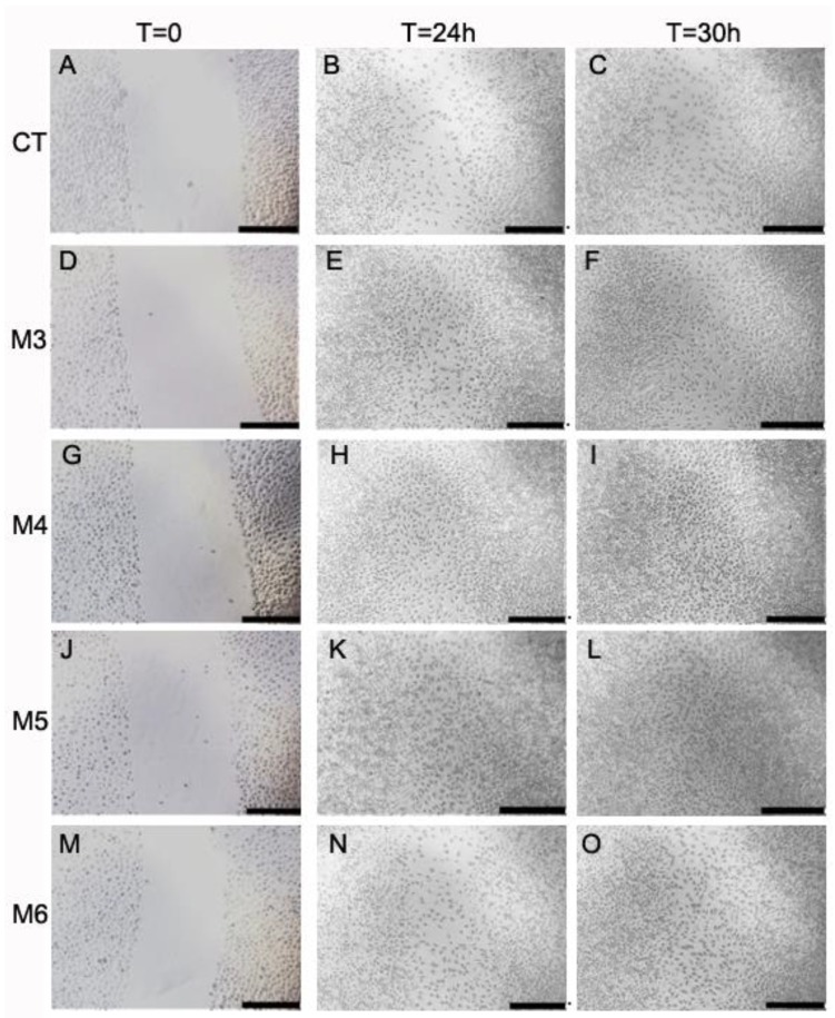
Figure 9.
Wound-healing assay in MCH-treated L929 fibroblasts. (A–O) Microphotographs taken at 0 h, 24 h, and 30 h with a 4× objective of L929 fibroblast monolayers during the wound-healing assay in the presence or absence of 50 µg/mL MCH fractions, in the area of the scratch made at time = 0. A–C control cells, D–F M3-treated cells, G–I M4-treated cells, J–L M5-treated cells, and M–O M6-treated cells. Black bars span 50 µm.

