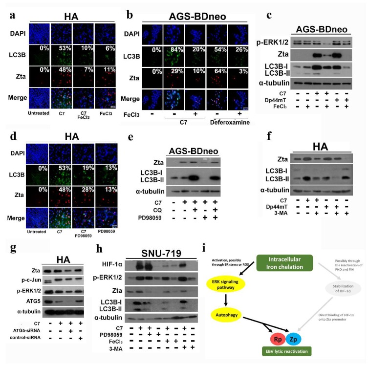
Figure 5.
C7 and iron chelators reactivate EBV lytic cycle via intracellular iron chelation and activation of the ERK-autophagy axis. (a) HA cells were incubated with either 20 μM C7 or 20 μM iron-precomplexed C7 for 48 h. Expression of Zta (red signals) and LC3B (green signals) was analyzed by immunofluorescence staining. DAPI (blue signals) stained cell nuclei. Scale Bar: 250 μm. (b) AGS-BDneo cells were incubated with either 20 μM C7, 20 μM iron-precomplexed C7, 1000 μM deferoxamine, or 1000 μM iron-precomplexed deferoxamine for 48 h. Expression of Zta (red signals) and LC3B (green signals) was analyzed by immunofluorescent staining. DAPI (blue signals) stained cell nuclei. Scale Bar: 250 μm. (c) AGS-BDneo cells were treated with either 20 μM C7, 20 μM iron-precomplexed C7, 20 μM Dp44mT, or 20 μM iron-precomplexed Dp44mT for 48 h. The expression of Zta and LC3B was analyzed by Western blotting. (d) HA cells were incubated with either 20 μM C7 or 20 μM C7 in combination with 50 μM PD98059 (MEK inhibitor) for 48 h. Expression of Zta (red signals) and LC3B (green signals) was analyzed by immunofluorescent staining. DAPI (blue signals) stained cell nuclei. Scale Bar: 250 μm. (e) AGS-BDneo cells were treated with either 20 μM C7, 20 μM C7 in combination with 10 μM chloroquine, 20 μM C7 in combination with 50 μM PD98059 (MEK inhibitor), 20 μM C7 in combination with 50 μM PD98059 (MEK inhibitor), or 10 μM chloroquine for 48 h. The expression of Zta and LC3B was analyzed by Western blotting. (f) HA cells were treated with either 20 μM C7, 20 μM C7 in combination with 5mM 3-MA, or 20 μM Dp44mT or 20 μM Dp44mT in combination with 5 mM 3-MA for 48 h. The expression of Zta and LC3B was analyzed by Western blotting. (g) Wild-type, atg5 knockdown and scramble control knockdown HA cells were treated with 20 μM C7 for 48 h. The expression of phosphorylated-c-Jun, phosphorylated-ERK1/2, ATG5, and Zta was analyzed by Western blotting. (h) SNU-719 cells were treated with either 20 μM C7, 20 μM C7 in combination with 50 μM PD98059, 50 μM PD98059 alone, 20 μM iron-precomplexed C7, 20 μM C7 in combination with 3-MA, or 3-MA alone for 48 h. The expression of HIF-1α, p-ERK1/2, Zta, and LC3B was analyzed by Western blotting. (i) Schematic illustration of EBV lytic reactivation via the proposed intracellular iron chelation-ERK-autophagy axis.

