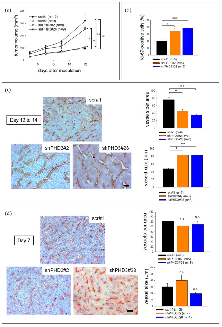
Figure 2.
PHD3 silencing leads to accelerated tumor growth, reduced tumor vessel density and enlargement of tumor vessels. (a) PHD3-deficient LM8 or control clones were injected subcutaneously into C3H mice. Tumor size measurement was performed every 2 to 3 days. The error bars represent mean values ± SEM. (* p < 0.05, ** p < 0.01). (b) Tumor sections were stained for Ki-67 and with dapi. The number of Ki-67-positive nuclei was determined. (* p < 0.05, *** p < 0.001). (c) PECAM and hematoxylin staining was performed on tumor sections. To determine the vessel density, the number of vessels in a given area was counted. The vessel size was also measured. The error bars represent mean values ± SEM. (* p < 0.05, ** p < 0.01). Scale bar: 150 µm. (d) ShPHD3 and control clones were injected subcutaneously into mice and the tumor size was measured. The experiment was terminated at day 7 after inoculation. To characterize the tumor vessels, PECAM and hematoxylin staining on tumor sections was performed. The error bars represent mean values ± SEM. Scale bar: 150 µm.

