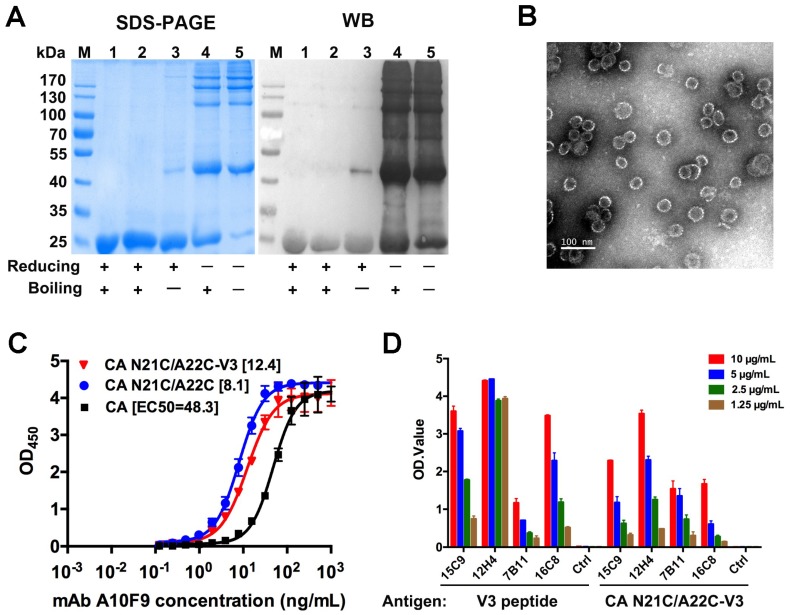
Figure 4.
Characterization of CA N21C/A22C-V3. (A) SDS-PAGE and western blotting of the assembled CA N21C/A22C-V3. Lane M, molecular weight marker; lane 1, protein before assembly; lane 2-5, protein after assembly. The primary antibody is a CA specific mAb A10F9. (B) Negative-stained electron microscopy image of a CA N21C/A22C-V3 particle. Scalebar is 200 nm. (C) Antigenicity of CA N21C/A22C-V3 was determined with the A10F9 mAb using indirect ELISA. Data were analyzed by GraphPad Prism software. The 50% maximal effective concentration (EC50) was calculated by a four-parameter logistic fit, and is indicated in the square brackets. (D) Antigenicity of CA N21C/A22C-V3 particles was measured by four anti-V3 mAbs (15C9, 12H4, 7B11 and 16C8) using indirect ELISA. These mAbs were found to have specific reactivity with the NL4-3 V3 synthesized peptide. CA N21C/A22C-V3 shows good reactivities with the mAbs, indicating that the V3 loop is properly displayed on the CA N21C/A22C particle scaffold. Data are the mean and standard deviations. All experiments were repeated three times.

