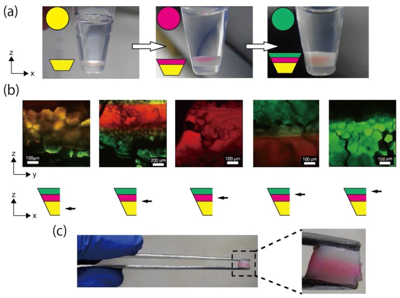
Figure 5.
Fabrication and observation of the triple-layered honeycomb microhydrogel network. (a) Fabrication process of the triple-layered honeycomb microhydrogel network. Initially, the first layer was formed at the bottom of the sampling tube (left). Then, the second layer was formed on the first layer (middle). Finally, the third layer was formed on the second layer (right). All layers were fabricated under FG = 1183× g. Upper insets: fluorescence coloring of the layers. Lower insets: simplified diagram of the layered honeycomb microhydrogel network. (b) CLMS images of the zy-plane of the cut triple-layered honeycomb microhydrogel network and illustrations of the observed position. The images in the upper panel are CLMS images of the zy-plane of the triple-layered structure. The observed position in the triple-layered structure is shown by the black arrows in the illustrations of the lower panel. (c) Direct handling of the triple-layered honeycomb microhydrogel network. The network was easily handled by a tweezer.

