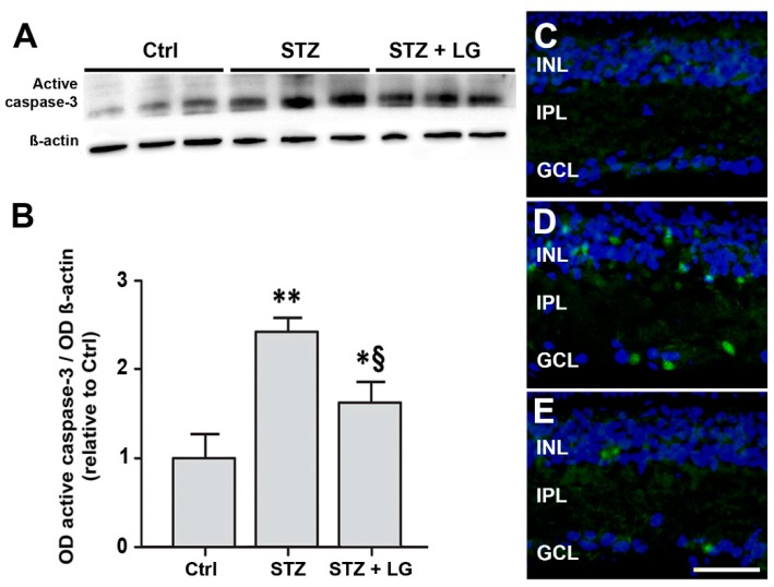
Figure 7.
(A) Western blots showing immunoreactive bands of active caspase-3 and of ß-actin, used as an internal standard, in control rat retinas and in retinas of STZ rats with or without LG treatment. (B) Quantitative analysis of the OD of the immunoreactive bands. Each column represents mean ± SD. * p < 0.05 vs. Ctrl; ** p < 0.01 vs. Ctrl; § p < 0.05 vs. STZ; n = 3 for all measures. Power value: 0.99. (C–E) Representative immunofluorescence images showing caspase-3 immunopositive cells in retinas of control rats (C), STZ rats (D), and STZ rats treated with LG (E). DAPI counterstain. Scale bar, 50 µm.

