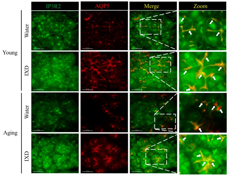
Figure 5.
Expression of IP3R2 and Aquaporin-5 (AQP5) in submandibular glands from young and aging rats treated with water or IXD extract (100 mg/kg) was observed by confocal microscopy. Left panel shows the IP3R2 expression in young and aging rats treated as mentioned in the figure. Second panel show the AQP5 staining within apical region of the acinar cells. Third panel show the overlay of IP3R2 and AQP5. Right panel show the zoomed picture of the co-localized IP3R2 and AQP5. White arrows indicate the overlay of IP3R2 and AQP5 within apical regions of submandibular gland acinar cells. The young group shows a uniform and clear co-localization of IP3R2 and AQP5, aging control rats show less overlapping, and IXD treated aging rats show the increased expression and higher overlapping in the apical regions. Scale bar: 25 microns.

