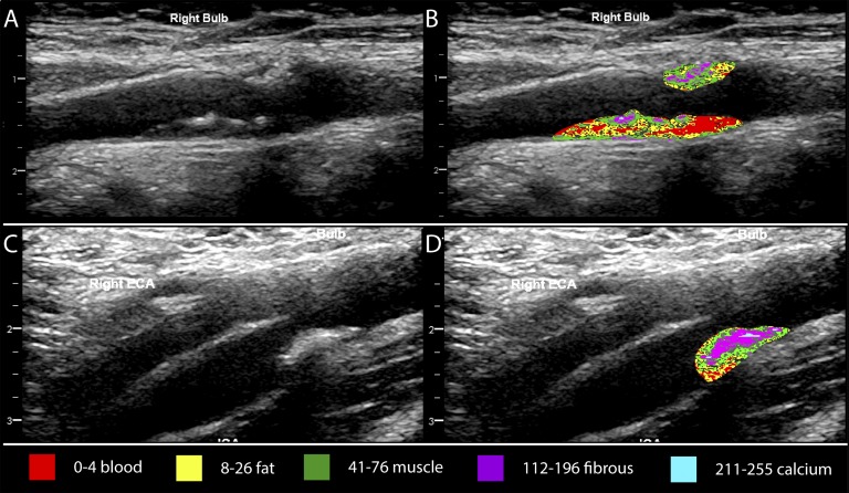
Figure 2.
Ultrasound grayscale color mapping of carotid plaque. (A, B) Right carotid bulb longitudinal view of plaque from a 66-year-old man. Phosphate level was 1.65 mmol/L, and FGF-23 was 94.4 RU/mL. Grayscale color mapping indicates that the plaque had a higher percentage of pixels in the blood (red) and fat-like (yellow) tissue ranges. (C, D) Right carotid bulb longitudinal view of plaque from a 68-year-old man. Phosphate level was 1.25 mmol/L, and FGF-23 was 71.9 RU/mL. Grayscale color mapping indicates that the plaque had a higher percentage of pixels in the fibrous-like tissue range (purple) with some calcification (blue). ECA, external carotid artery.

