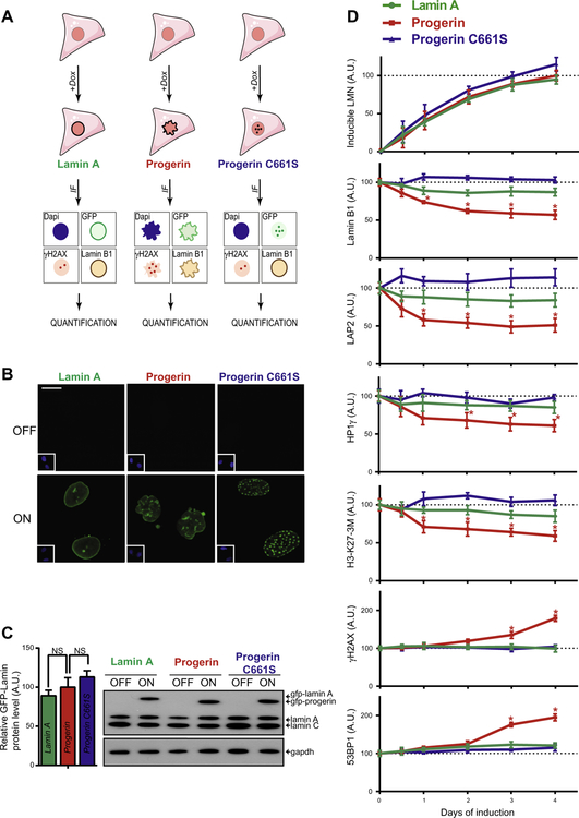
Fig. 1.
Characterization of an inducible GFP-progerin fibroblast cell system. (A) Schematic representation of stable human dermal fibroblasts cell lines containing doxycycline-inducible GFP-tagged lamin A, progerin or progerin C661S mutant for use in immunofluorescence (IF) detection of GFP, lamin B1, γH2AX, and DNA DAPI stain. (B) Representative IF images of DNA DAPI stain (inset bottom left) and GFP signal of all three GFP-lamin inducible fibroblasts cell lines in the absence and presence of doxycycline for 4 days. Scale bar: 10 μM. (C) Western blot for uninduced and 4 days induced GFP-lamin fibroblast cell lines and quantification of β-actin normalized lamin protein levels. Values represent averages ± SD from 3 experiments. (p > 0.05). (D) Quantification of IF signals (see Section 2) of induced GFP-lamin protein levels as well as lamin B1, LAP2, HP1γ, tri-methylated lysine 27 on histone 3 (H3-K27–3M), γH2AX and 53BP1 HGPS markers. Values represent averages ± SD from 3 experiments (N > 300, *p < 0.05 between GFP-lamin A and GFP-progerin cell lines).

