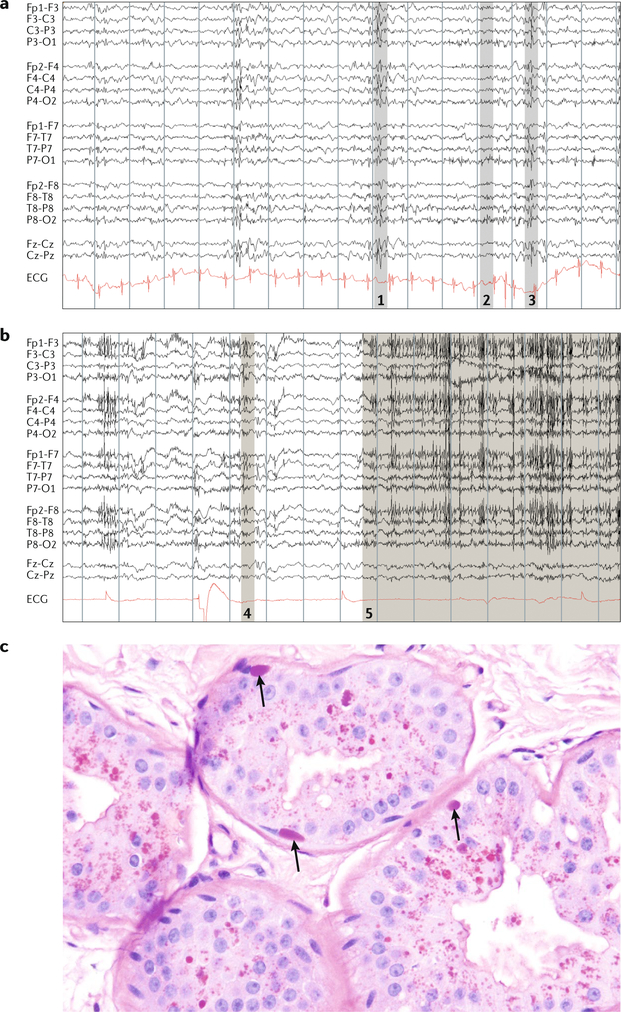
Fig. 4 |. Diagnosis of Lafora disease.
a | A 16 s EEG trace in an awake patient with Lafora disease (compound heterozygous for EPM2A: Arg241X and Pro211Leu) at age 20 years, 5 years into the disease. Note the slow background rhythms and generalized irregular spike-wave discharges associated with myoclonus. At this age, the patient was still conversant, but each thought and sentence was interrupted by myoclonic absences. b | EEG in the same patient at age 28 years in an unresponsive wakeful state. Grey shaded areas in parts a and b denote patient movements: 1, arm jerk; 2, head jerk; 3, head and arm jerk; 4, twitch; and 5, eyes upward and blinking. c | Skin biopsy in the same patient at age 20 years. Arrows indicate Lafora bodies in the myoepithelium of apocrine glands.The equally stained structures near the lumina of the glands are not Lafora bodies but the normal secretory materials of these cells. Owing to a lack of experience with the disease, many laboratories mistake these materials for Lafora bodies, leading to false-positive diagnoses. ECG, electrocardiogram

