
Fig. 1.
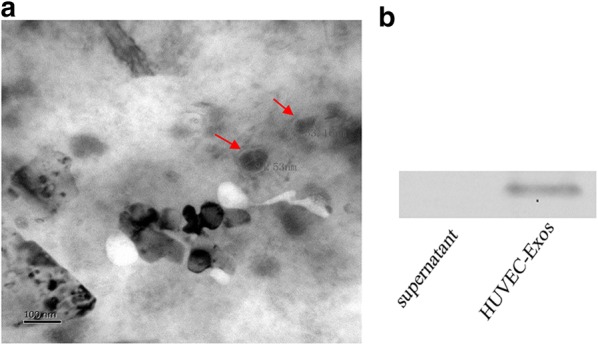
Identification of HUVEC-Exos. The HUVECs were cultured for 48 h in NG (5 mmol/l) or HG (30 mmol/l) conditions. a Electron microscopic image of HUVEC-Exos. b Western blot of exosomal-specific marker CD63 in HUVEC-Exos. Scale bar, 100 nm
Official websites use .gov
A
.gov website belongs to an official
government organization in the United States.
Secure .gov websites use HTTPS
A lock (
) or https:// means you've safely
connected to the .gov website. Share sensitive
information only on official, secure websites.
Identification of HUVEC-Exos. The HUVECs were cultured for 48 h in NG (5 mmol/l) or HG (30 mmol/l) conditions. a Electron microscopic image of HUVEC-Exos. b Western blot of exosomal-specific marker CD63 in HUVEC-Exos. Scale bar, 100 nm