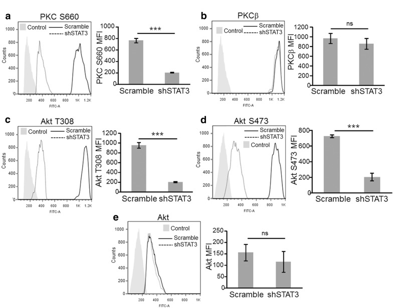
Fig. 4.
Flow Cytometry analysis of total and activated PKC and Akt upon STAT3 silencing. a Flow cytometry analysis of P-PKC a, PKCβ b, P-Akt (T308) c, P-Akt (S473) d and Akt e expression. Cells were collected and stained as described in “Methods”. Light-grey filled histogram correspond to control Ab and open histograms to the corresponding specific abs. Solid and broken lines correspond to scramble and shSTAT3 cells, respectively. Bar graphs show the mean mean fluorescence intensity (MFI) ± S.D. (from three independent experiments) for the indicated antibody. Statistical significance was tested by a one-tailed Student’s T-Test, ***p < 0.0001, ns: not significant, n = 3

