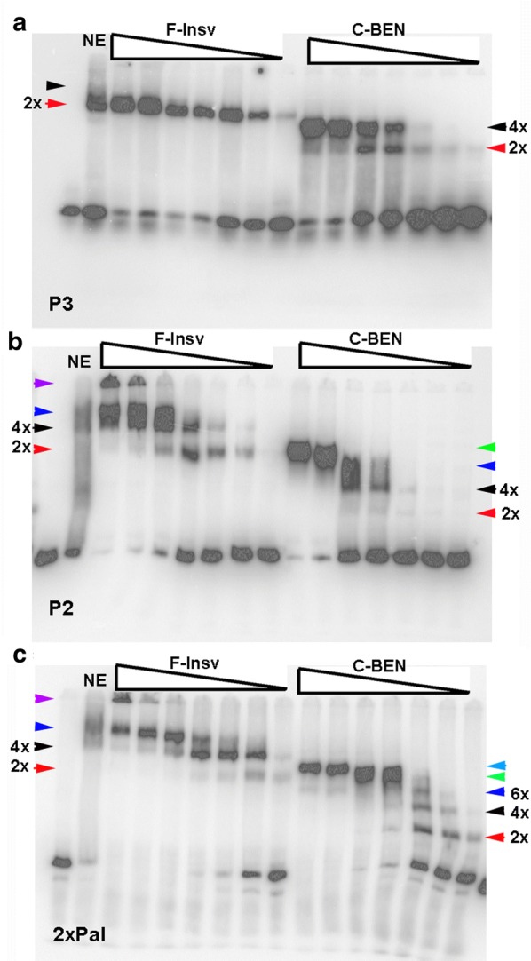
Fig. 4.

HT:F-Insv and HS:C-BEN generate complex patterns of shifts with different substrates. a HT:F-Insv and HS:C-BEN shifts of probe P3. b probe P2 (see diagram in Fig. 1). c probe 2xPal (see diagram in Fig. 1). Predicted multimers are indicated by arrowheads (see also text). Amounts of protein added (left to right) to the reaction mix were estimated based on the relative intensity of the Coomassie-stained protein bands in SDS-PAGE gels: 0.5 μM, 0.25 μM, 0.05 μM, 0.025 μM, 0.005 μM, 0.0025 μM, and 0.0005 μM
