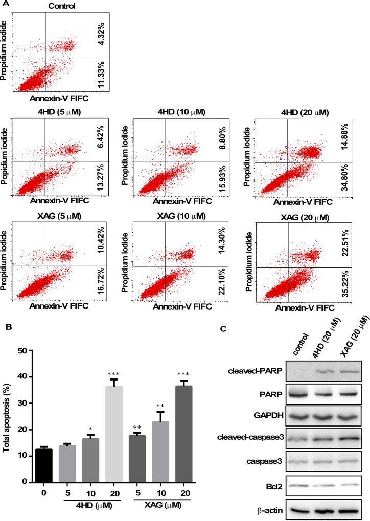
Figure 4. 4HD or XAG induces apoptosis in melanoma cells.
(A and B) SK-MEL-28 cells (2.5 × 10⁵/well) were incubated with 4HD or XAG (5, 10, or 20 µM), or vehicle control for 72 h. Cells were collected and apoptosis was detected using flow cytometry and Annexin V staining. Data are represented as mean values ± S.D. as determined from 3 independent experiments and the asterisks indicate a significant decrease compared with untreated control cells (*, p < 0.05; **, p < 0.01; ***, p < 0.001). (C) The cells were incubated with 20 µM 4HD or XAG or vehicle control for 72 h, then the effect of 4HD or XAG on apoptosis-associated protein expression was determined by western blotting.

