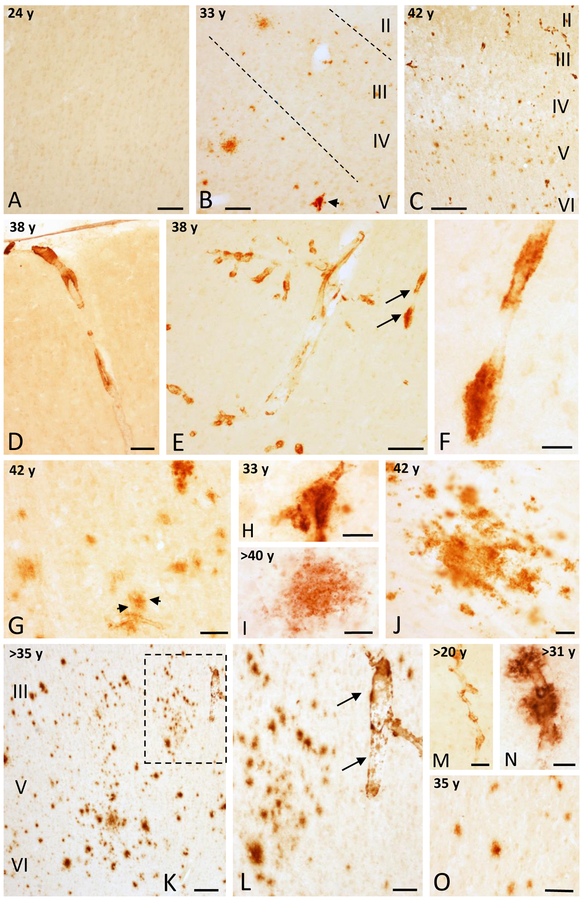
Figure 1.
Photomicrographs showing the absence of APP/Aβ-ir profiles in area 9 in a 24 year-old female gorilla (A) and the presence of some APP/Aβ-ir profiles in a 33 (B) and many more in a 42 (C) year-old female gorilla. Images showing variation in APP/Aβ-ir blood vessels (D-F) in the area 9 in a 38 year-old female gorilla. F. High-magnification photomicrograph of APP/Aβ-positive blood vessel (arrows) shown in panel E. G and H. Aβ positive plaque (black arrows) associated to a blood vessel in a 42 year-old gorilla (G), and blood vessel with Aβ accumulations from panel B (arrow) in a 33 year-old female gorilla (H). I and J. High-magnification photomicrographs showing a diffuse APP/Aβ-ir plaques in the frontal cortex of a >40 (I) and a 42 (J) year-old female gorilla. K. Photomicrograph illustrating numerous APP/Aβ-positive plaques in the posterior orbital cortex (area 44) in a >35 year-old female gorilla. L. High-magnification photomicrograph of the boxed area in K showing APP/Aβ-positive plaques and an adjacent blood vessel (arrows). M and N. APP/Aβ-positive parenchymal blood vessels in a >20 and >31 year-old male gorillas, respectively. Note the Aβ accumulations in the blood vessel in >31 year-old male gorilla. O. APP/Aβ-positive plaques in a 35 year-old male gorilla. Abbreviations: I, II, III, IV, V and VI, cortical layers. Scale bars = 200 μm in K; 100 μm in B-E and O; 75 μm in L; 50 μm in A, G, and M; 25 μm in F, H-J, and N.

