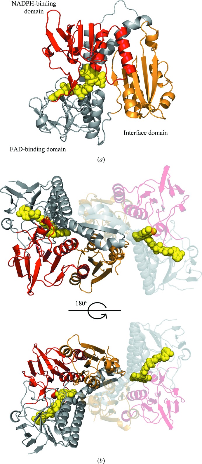
Figure 2.
Structure of the glutathione reductase from S. pneumoniae. (a) The structure of the SpGR monomer is shown in cartoon representation with the FAD-binding domain in grey, the NADPH-binding domain in red and the interface domain in gold. The FAD cofactor is represented as yellow spheres. (b) Structure of the SpGR dimer: both monomers are coloured according to their domain structures as in (a).

