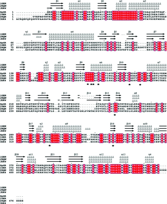
Figure 4.
Sequence alignment of SpGR against the EcGR, hGR and yGR homologues. The alignment was performed using T-Coffee and ESPript (http://tcoffee.crg.cat/apps/tcoffee/do:expresso; Robert & Gouet, 2014 ▸). The secondary-structural elements were identified from PDB entries 1ger, 2hqm and 3dk4 using ESPript and are displayed at the top of the alignment. The α-helices and β-sheets are denoted α and β, respectively. Conserved residues are indicated by white lettering on a red background. Resides that form the NADPH-binding site are indicated with black stars.

