Figure 3.
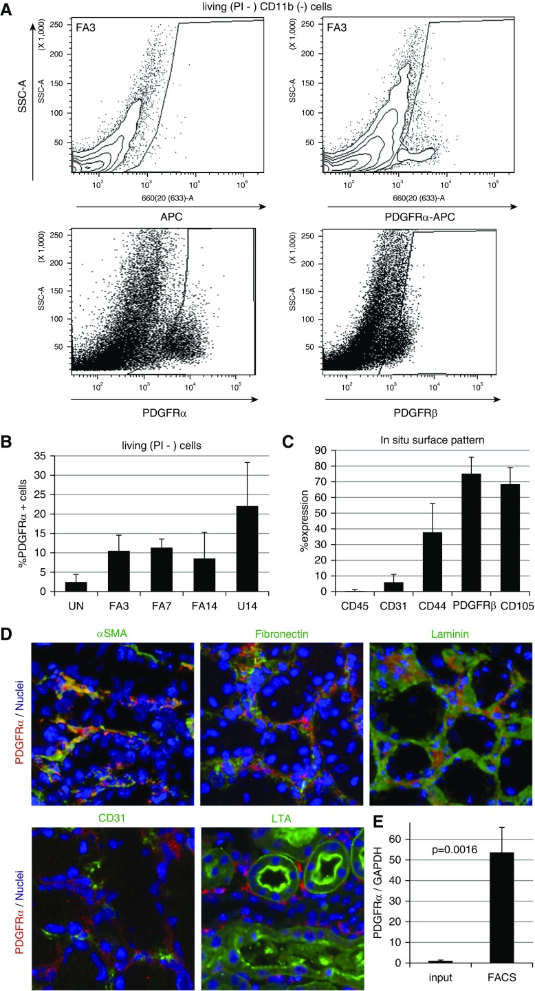
PDGFrα-positive cells can be sorted with purity and specificity from adult kidneys. (A) Representative flow cytometry plot for sorting PDGFRα-positive cells from whole adult kidney 3 days post-FA, using either a control (left panel) or anti-PDGFRα mAb (APA5, right panel). Cells in gated area were selected. (B) The percentage of PDGFRα-positive cells from whole kidneys sorted from each disease model and uninjured control kidneys (no injury, n=7; FA3, n=9, FA7, n=5; FA14, n=5; U14, n=4). (C) Flow cytometry analysis shows that PDGFRα-sorted cells include pericyte/MSC marker–positive cells (CD44, PDGFRβ, and CD105), but not endothelial (CD31) and hematopoietic (CD45) cell populations (n=3). (D) Immunostaining demonstrates that anti-PDGFRα mAb (APA5) recognizes cells specifically confined to the tubulointerstitial compartment. In FA3 kidney, PDGFRα-positive (red) cells stain positive for αSMA, overlap with ECM (fibronectin and laminin), but do not costain with endothelial marker (CD31) and proximal tubular marker (LTL-lectin). (E) Quantitative RT-PCR analyses of PDGFRα mRNA from whole FA3-treated kidneys and PDGFRα-sorted cells show approximately 50-fold enrichment (n=3). SSC-A, Side scatter; UN, Uninjured.

