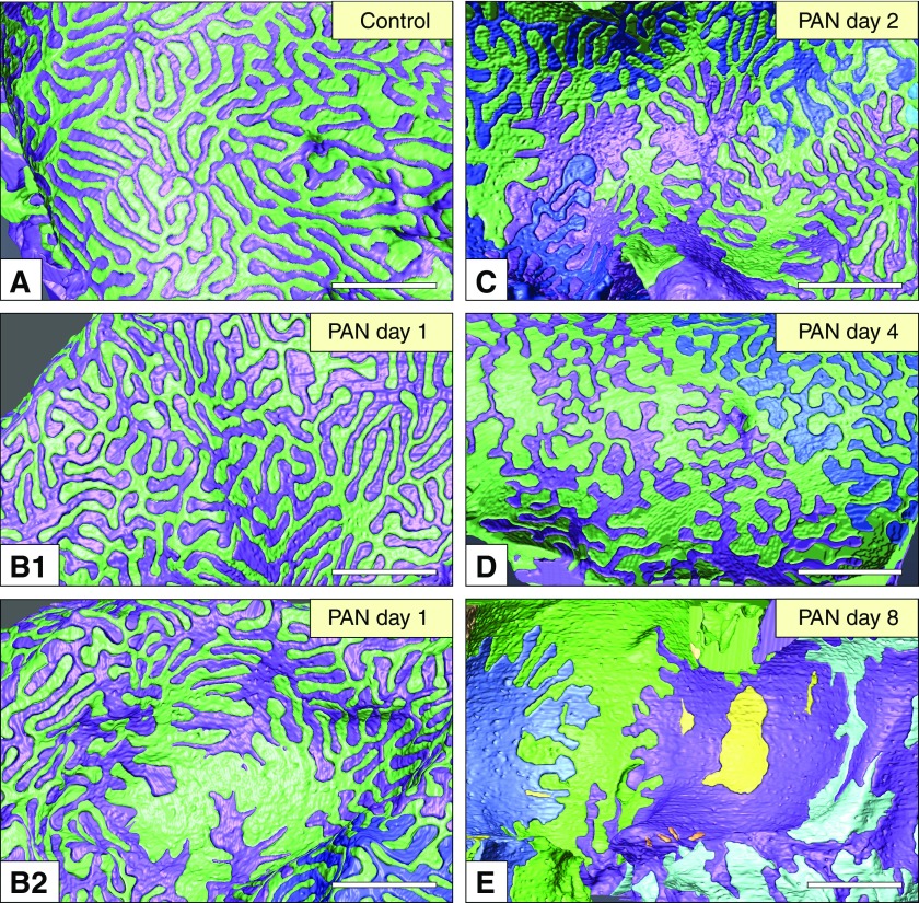
Figure 2.
Reconstruction of podocytes based on FIB/SEM tomography is useful for observing the alterations in the basal surface structure of podocytes. Individual podocytes are shown in different colors. The basal view of the reconstructed podocytes is useful to analyze structural alterations in the foot processes. (A) In healthy (control) podocytes, foot processes exhibited a uniform width. (B1 and B2) PAN nephrotic podocytes lost this uniformity in the foot processes from day 1. The uniformity in width was further lost on (C) days 2 and (D) 4. (E) The podocytes formed a large adhesive surface on day 8, although short interdigitating processes remained. The yellow masses represent the CFs of podocyte (E), which are frequently found in the PAN nephrotic glomeruli. Scale bar, 2 μm.

