Figure 6.
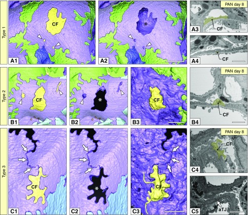
Podocyte fragmentation progressed in association with foot process effacement. Three kinds of podocyte CFs were recognized at least. (A1, A2, B1, B2, C1, and C2) Basal view. The CFs are represented as yellow masses. (A2, B2, and C2) The CFs were removed to show their positions in relation to the neighboring purple podocyte. Asterisks indicate the space for CF. (B3 and C3) Luminal view. (A1 and A2) Type-1 CFs. A large CF (CF in A1) was completely covered by a deformed purple podocyte. Arrowheads indicate two small type-1 CFs. (B1–B3) Type-2 CFs. A large CF (CF in B1) penetrated the deformed purple podocyte. Arrowheads indicate two small type-1 CFs. (C1–C3) Type-3 CFs. CF surrounded by two deformed primary processes of purple podocyte, whose distal ends were connected by an aTJ (arrows). (A3, A4, B4, and C4) FIB/SEM images of CFs shown in (A1, B1, and C1). (C5) FIB/SEM image of aTJ in purple podocyte (arrow). Scale bars, 1 μm.

