Figure 7.
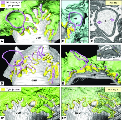
Reconstruction of podocytes based on FIB/SEM tomography is useful for observing the alterations in the junctional structure. (A–F) Day 4. (A, B, D, and E) Luminal view. The slit diaphragm (pink) and TJ (yellow) were put on the reconstructed green podocytes. In some parts, the slit diaphragm coexisted on the TJ (arrowheads in E). Part of the slit diaphragm moved further toward the luminal side without associating with the TJ and held its position as an eSD between a pair of cup-shaped protrusions (asterisks in A, B, and E) derived from two neighboring primary processes. (C) FIB/SEM image of the cup-shaped protrusions (asterisk) linked by eSDs (arrows). (D) To clearly show the junctional apparatus, the green podocyte was removed from (A). (F) FIB/SEM image of the slit diaphragm (pink arrowheads) coexisting with the TJ (yellow arrowheads). (G1 and G2) Day 8. Luminal view. The podocytes were predominantly connected to each other by the TJs (yellow). Scale bars, 200 nm in (C and F); 500 nm in (A, B, D, E, and F2)

