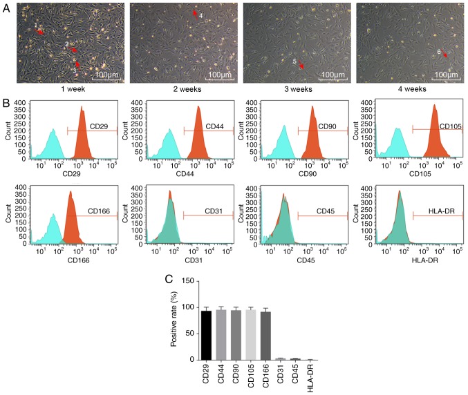
Figure 1.
Morphological changes and surface markers of ADSCs. (A) Representative images of the morphological changes of ADSCs during 1, 2, 3 and 4 weeks (magnification, ×100), as observed with an inverted light microscope. Arrow 1 represents multinucleate cells; arrow 2 represents long spindle cells; arrow 3 represents round cells; arrow 4 represents polygonal cells; arrow 5 represents myotube cells; and arrow 6 represents star-shaped cells. (B) Distribution of surface markers of ADSCs, as detected by flow cytometry. (C) Quantification of % positive rates of ADSC surface markers. ADSCs, adipose-derived stem cells.

