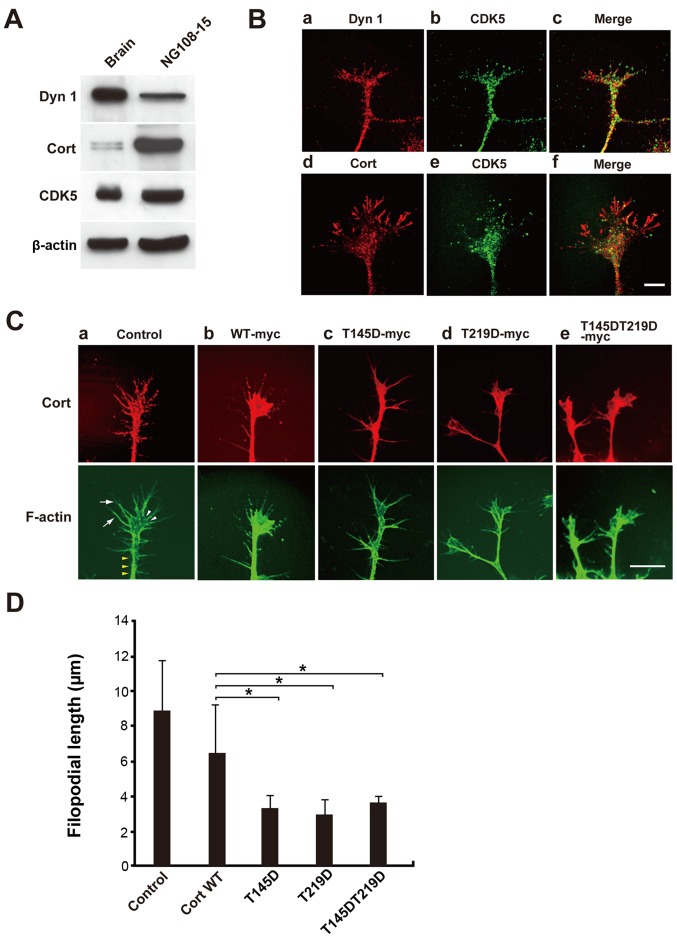
Figure 4.
NG108-15 cells expressing phosphomimetic Cort mutants exhibit aberrant lamellipodia and short filopodia. (A) Expression of Dyn 1, Cort and CDK5 in NG108-15 cell lysates was confirmed by western blotting. In total, 10 µg of mouse brain homogenate or 20 µg of NG108-15 cell lysate was loaded per lane. (B) Immunofluorescence analysis showing that endogenous Dyn 1, Cort and CDK5 partially colocalized in lamellipodia and filopodia of NG108-15 cells. Scale bar, 10 µm. (C) NG108-15 cells were not transfected (a; control) or transfected with c-myc-tagged (b) WT Cort or the (c) T145D, (d) T219D or (e) T145DT219D mutant, fixed, and stained with a monoclonal anti-c-myc antibody (red). F-actin was stained with Alexa Fluor 488-conjugated phalloidin (green). (Ca) Typical cellular protrusion (yellow arrow heads), lamellipodia (white arrow heads) and filopodia (white arrows) are presented. Filipodia were markedly shorter in T145D-, T219D- and T145DT219D-expressing cells than in WT Cort-expressing cells. Scale bar, 10 µm. (D) Morphometric analysis of filopodial length in (C). Data are presented as the mean ± standard error of the mean of 33-60 filopodia per experimental condition (n=3 independent experiments; *P<0.05). Cort, cortactin; Dyn 1, dynamin 1; CDK5, cyclin-dependent kinase 5; WT, wild-type.

