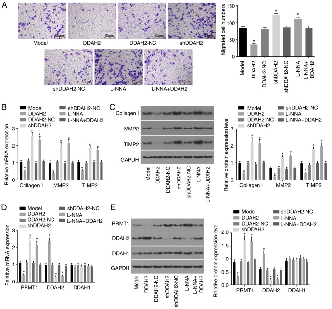
Figure 4.
DDAH2 inhibits myocardial cell migration. (A) Migration ability of H9C2 myocardial cells under high glucose conditions following 48 h of treatment with lentivirus expressing DDAH2 or shDDAH2, and/or L-NNA (magnification, ×200). (B) mRNA levels of collagen I, MMP2 and TIMP in H9C2 myocardial cells under high glucose conditions following 48 h of treatment with lentivirus expressing DDAH2 or shDDAH2, and/or L-NNA. (C) Protein levels of collagen I, MMP2 and TIMP in H9C2 myocardial cells under high glucose conditions following 48 h of treatment with lentivirus expressing DDAH2 or shDDAH2, and/or L-NNA. (D) mRNA expression of PRMT1, DDAH2 and DDAH1 in H9C2 myocardial cells under high glucose condition following 48 h of treatment with lentivirus expressing DDAH2 or shDDAH2, and/or L-NNA (E) Protein expression of PRMT1, DDAH2 and DDAH1 in H9C2 myocardial cells under high glucose conditions following 48 h of treatment with lentivirus expressing DDAH2 or shDDAH2, and/or L-NNA. Data are presented as the mean ± standard deviation and analyzed by one-way analysis of variance. All data are representative of three independent experiments. *P<0.05, vs. model group. DDAH, dimethylarginine dimethylaminohydrolase; PRMT1, protein arginine N-methyltransferase 1; NC, negative control; sh, short hairpin RNA; MMP2, matrix metalloproteinase 2; TIMP2, tissue inhibitor of metalloproteinase 2; GAPDH, glyceraldehyde-3-phosphate dehydrogenase.

