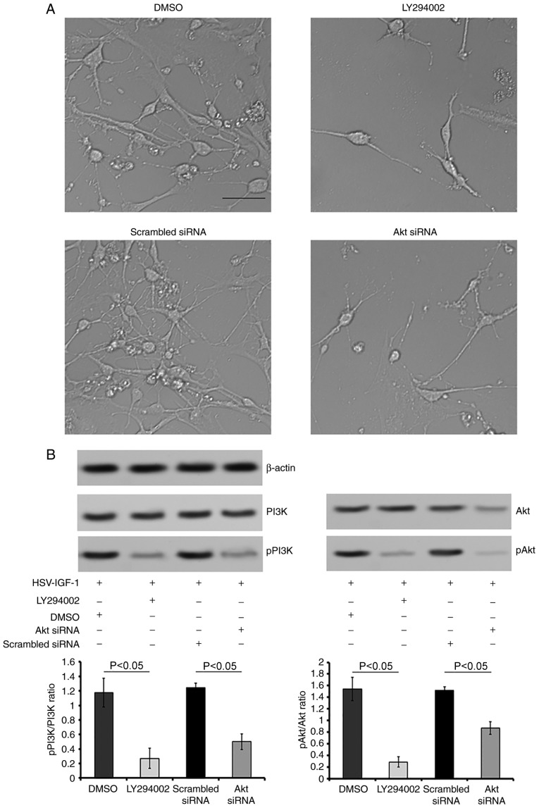
Figure 8.
Roles of PI3K/Akt in IGF-1 induced neuroprotection in vitro. (A) Morphological changes of cultured DRG neurons following treatment with DMSO (control), LY294002, scrambled siRNA (control) and Akt siRNA in cultured DRG neurons transfected with HSV-IGF-1 (magnification, ×200). (B) Representative blots of PI3K, pPI3K, Akt and pAkt proteins detected by western blot analysis. The ratios of pPI3K/PI3K and pAkt/Akt are also shown. Values are plotted as the mean ± standard deviation (n=5). IGF-1, insulin-like growth factor 1; DRG, dorsal root ganglia; siRNA, small interfering RNA; PI3K, phosphatidylinositol 3-kinase; pPI3K, phosphorylated PI3K; pAkt, phosphorylated Akt; DMSO, dimethyl sulfoxide.

