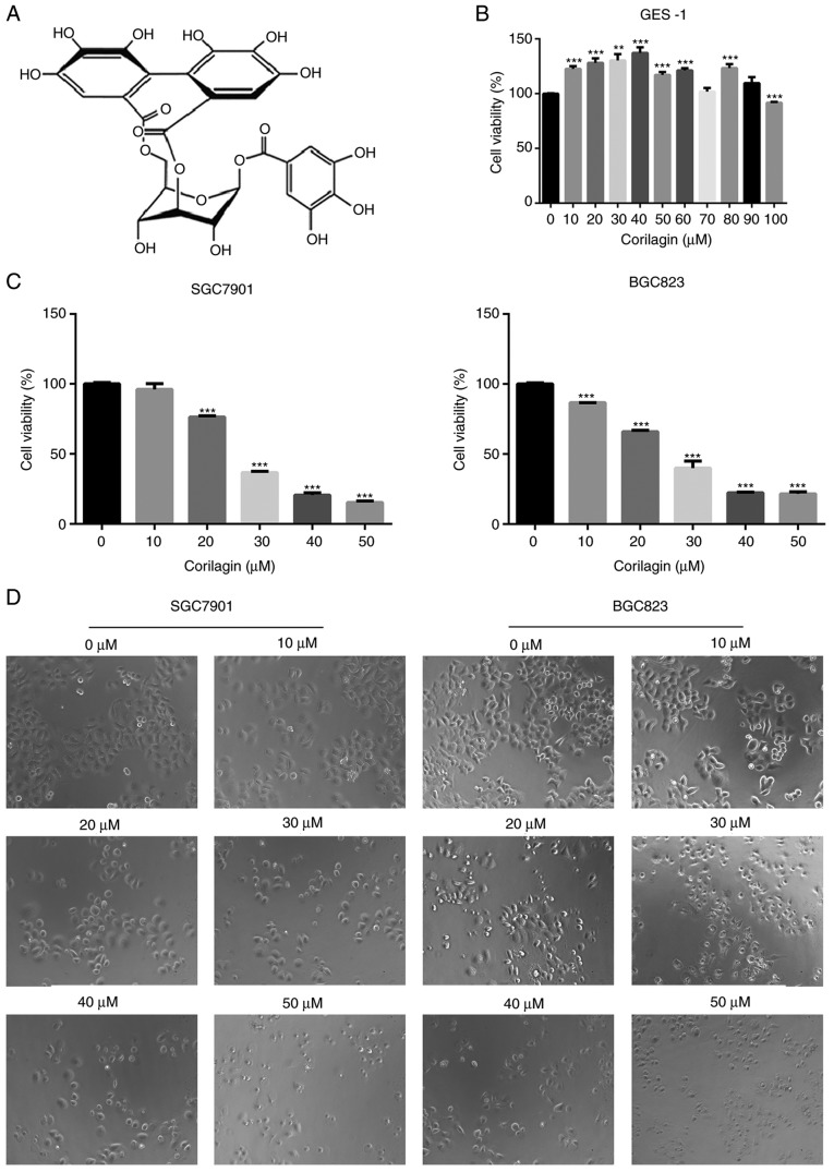
Figure 1.
Effect of corilagin on the viabilities of human gastric cancer cells. (A) Chemical structure of corilagin. (B) GES-1 cells were treated with various concentrations of corilagin (0-100 µM) for 24 h. (C) SGC7901 and BGC823 cells were treated with different concentrations of corilagin (0, 10, 20, 30, 40 and 50 µM) for 24 h. The cell viabilities were examined using a 3-(4,5-dimethylthiazol-2-yl)-2,5-diphenytetrazolium bromide assay. (D) SGC7901 and BGC823 cells were exposed to corilagin at different concentrations (0-50 µM) for 24 h, and images were captured by microscopy at magnification, ×200. Data are reported as the mean ± standard deviation (n≥3) of three replicate experiments. Significant differences from the control group were measured using Student's t-test. **P<0.01, ***P<0.001.

