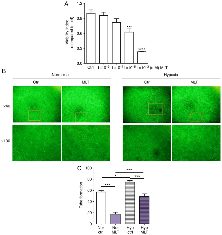
Figure 1.
MLT suppresses the viability and angiogenesis of HUVECs. (A) HUVECs were treated with MLT at various concentrations (1×10⁻⁹, 1×10⁻⁷, 1×10⁻⁵ or 1×10⁻³ mM) or left untreated for 24 h. The viability of HUVECs was determined using a Cell Counting kit 8 viability assay. (B) HUVECs were treated with MLT (1×10⁻⁵ M) under normoxia or hypoxia conditions, and imaged under an Olympus microscope. Magnification, ×40 or ×100. (C) The statistics histogram for. (B) Data are presented as the mean ± standard error of the mean. *P<0.05, ***P<0.001 or ****P<0.0001, compared with the Ctrl group. Ctrl, control; MLT, melatonin; HUVECs, human umbilical vein endothelial cells; Nor Ctrl, Ctrl HUVECs under normoxia condition; Nor MLT, MLT-treated HUVECs under normoxia condition; Hyp Ctrl, Ctrl HUVECs under hypoxia condition; Hyp MLT, MLT-treated HUVECs under hypoxia condition.

