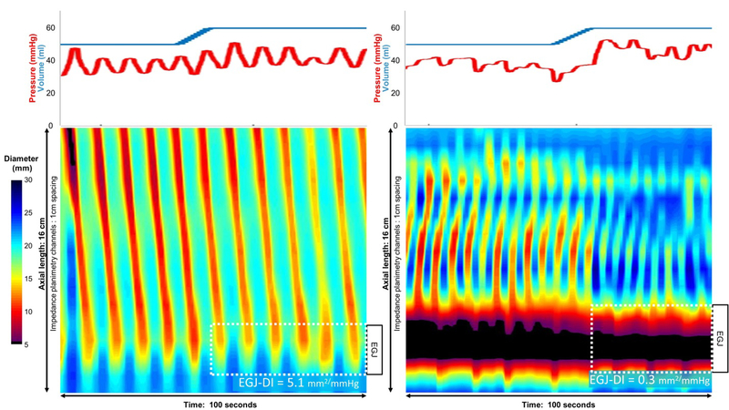
Figure 2.
Two images of FLIP Panometry showing repetitive antegrade cotractions (left) and repetitive retrograde contractions (right). The plots on top indicate the volume within the FLIP balloon (blue) and the corresponding pressure (red). On the topography plots time is on the x-axis, position along the 16 cm balloon on the y-axis, and spectral color indicates luminal diameter at each coordinate as per the scale. With the exception of the small blackened are on the first contraction in the left panel and the EGJ in the right panel, these are all non lumen-occluding contractions. Repetitive antegrade contractions are a normal finding and the patient on the left had normal motility on HRM. However, repetitive retrograde contractions are rarely found in normals and are usually indicative of obstructive physiology; the patient on the right had type III achalasia. The esophagogastric junction distensibility index (EGJ-DI) is measured at 60 ml distension with 2.8 mm2/mmHg being the lower limit of normal.

