Fig. 2.
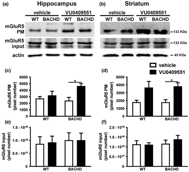
VU0409551 treatment increased mGluR5 cell surface expression in BACHD mice. Shown are representative immunoblots for mGluR5 cell surface (upper panel) and total cell lysate expression (middle panel), as well as representative immunoblots for actin (lower panel) in hippocampal (a) and striatal (b) slices of wild-type (WT) and BACHD mice, treated with either vehicle or VU0409551. Graphs show the densitometric analysis of the cell surface expression of mGluR5 in hippocampal (c) and striatal (d) slices, as well as mGluR5 total cell lysate expression in hippocampal (e) and striatal (f) slices of WT and BACHD mice, treated with either vehicle or VU0409551. Data represent the mean ± SEM. Number of mice: n = 4. ♦indicates significant difference (p < 0.05).

