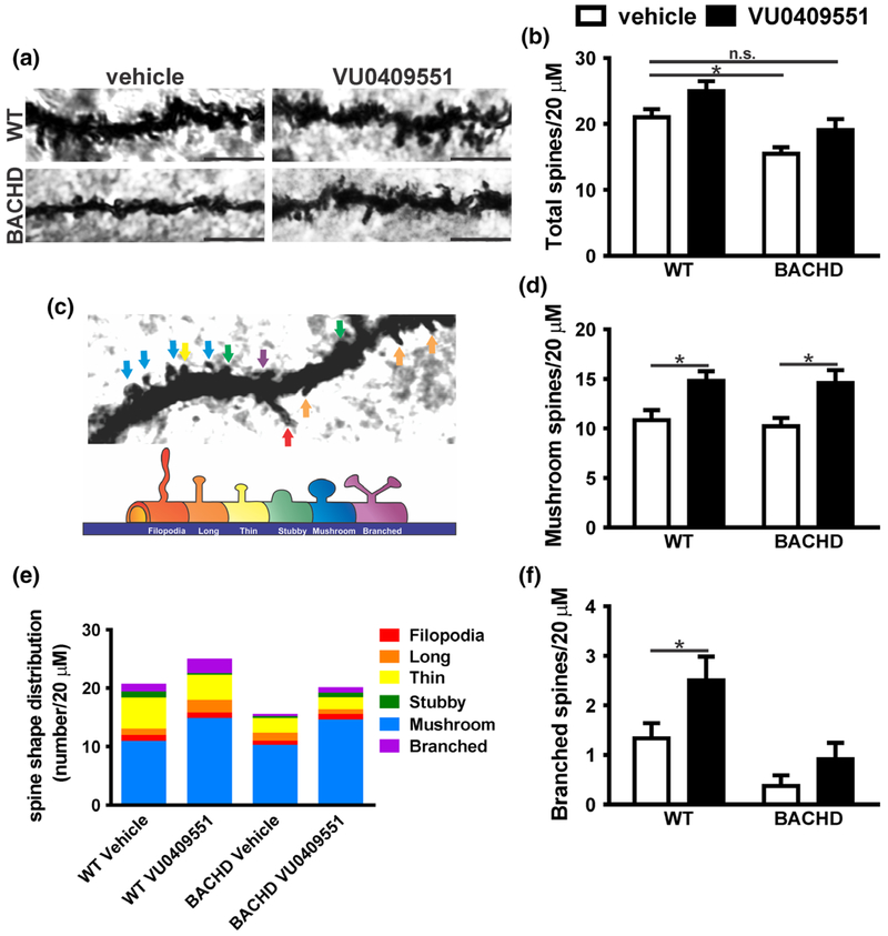
Fig. 5.
VU0409551 treatment increased dendritic spine maturation. (a) Shown are representative confocal micrographs from tertiary dendrites of hippocampal neurons from wild-type (WT) and BACHD mice, treated with either vehicle or VU0409551. Dendrite segments shown are 20 μm in length and size bar corresponds to 5 μm. (b) Graph shows dendritic spine density (total number of dendritic spines per 20 μm). (c) Upper image: Confocal image of a dendrite containing all possible shapes of dendritic spines, which are pointed by colored arrows. Lower image: Schematic drawing of spines shown in: red: filopodia; orange: long; yellow: thin; green: stubby; blue: mushroom; purple: branched. (e) Colored graph shows the contribution of each type of spine per 20 μm of dendrite segment. Graphs show the total number of mature mushroom (d) and branched (f) spines per 20 μm of dendrite segment. Data represent the mean ± SEM. Number of dendrites: WT-vehicle n = 15, WT-VU0409551 n = 12, BACHD-vehicle n = 12, and BACHD-VU0409551 n = 12. *Indicates significant difference (p < 0.05).

