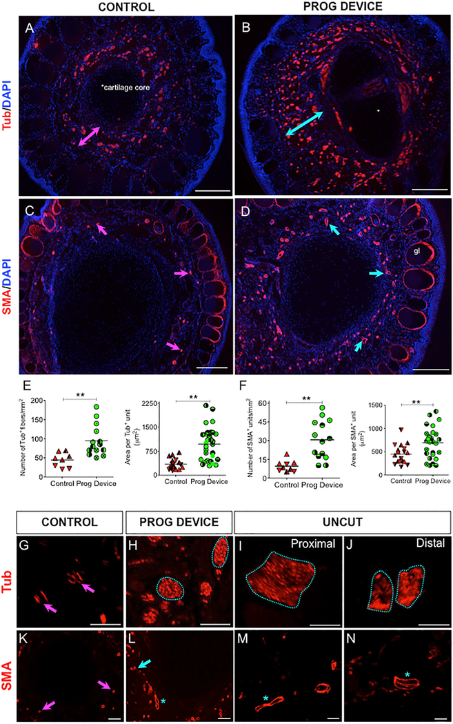
Figure 4. Prog-Device Treated Animals Show Significantly Greater Innervation and Vascularization than Untreated Animals at the Most Distal Part of the 9.5-mpa Regenerate, with Patterns Closer to Uncut (Intact) Limbs.
(A–D) Low-magnification immunofluorescence images of DAPI-counterstained cross-sections revealing innervation (anti-acetylated a-tubulin antibody [Tub]) and vascularization (anti-smooth-muscle actin antibody [SMA]) of amputated untreated (Ctrl) and amputated plus Prog-device (Prog-device) treated animals. The overall area occupied by Tub+ fibers is significantly greater in treated tips (turquoise double-headed arrow in B, compared to magenta double-headed arrow in A). Similarly, the density and extension of the blood vessels for Prog-device treated animals (D, turquoise arrows) is higher when compared to untreated animals (C, magenta arrows). gl, SMA+ epidermal glands were not included in the analysis. Scale bars, 250 μm. The cartilage core of the 9.5-mpa regenerates is indicated in (A). For a schematic representation of the plane cut, see igure S5A.
(E and F) Quantitative results of number of positive units per square millimeter and area per unit for Tub (E) and SMA (F) immunofluorescence in Ctrl (red) and Prog-device groups (green). Values are represented with scatterplots, in which each dot represents one histological section, and each dot style represents one animal (with at least three histological sections). Statistical analysis was performed on the pooled individual sections. Horizontal line indicates mean. p values after Mann-Whitney test (unequal variances) or t test (equal variances; only for E, left) are indicated as **p < 0.01.
(G–N) High-magnification images revealed that while in untreated animals, the typical nerve patterning is composed of individual and unpatterned nerve fibers (magenta arrows in G), Tub+ axons in Prog-device animals show a tendency to group organization, forming bundles (turquoise line-encircled areas in H), similar to the nerve organization in intact or uncut limbs (turquoise line-encircled areas in I and J). Both the number and morphology and area of the SMA+ vessels for Prog-device tips were clearly closer to the intact limb (turquoise arrow and asterisks in L, M, and N) than to the Ctrl untreated limbs (small vessels indicated by magenta arrows in K). See Table S4 for quantitative data. Scale bars, 100 μm.

