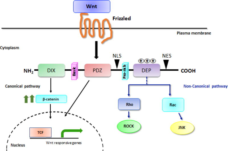
Figure 2. The structure of DVL proteins.
DVL is made up of three conserved motifs namely an amino-terminal DIX domain, a central PDZ, and a carboxyl-terminal DEP domain. In addition to these three domains, DVL harbors two regions with positively charged amino acid residues (basic and proline-rich domain) plus a nuclear import (NLS) and a nuclear export signal (NES). The DIX and PDZ domain relay signal to canonical pathway (marked in black arrows), whereas DEP domain mainly regulates membrane localization of DVL and propagates non-canonical pathway (marked in blue arrows).

