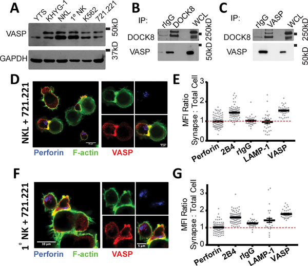
FIGURE 1.
VASP localizes to the immunological synapse and granules in NK cells. (A) Whole-cell lysates from the panel of cell lines indicated were immunoblotted for VASP and GAPDH. (B) DOCK8 and (C) VASP protein were immunoprecipitated from NKL lysates and immunoblotted for the presence of DOCK8 and VASP. Representative immunofluorescence images of NKL-721.221 (D) and primary NK-721.221 (F) conjugates stained for VASP (red), perforin (blue) and F-actin (green). Quantitation of the Mean Fluorescence Intensity (MFI) of various proteins and rIgG at the cytotoxic synapse in comparison to the MFI of the entire NKL (E) or primary NK cell (G). All results are representative of at least three independent experiments.

