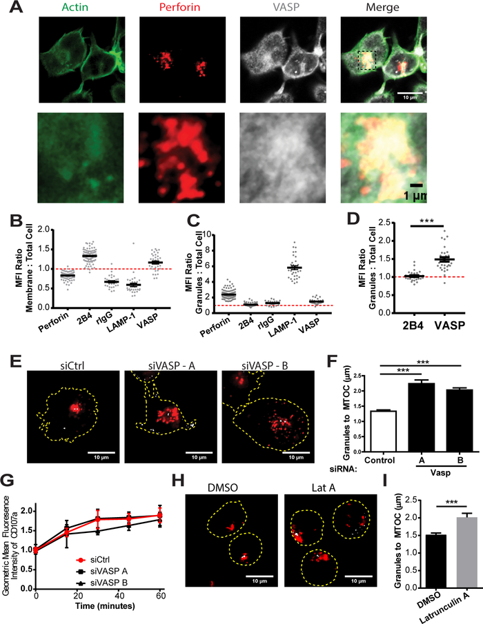
FIGURE 7.
Constitutive granule polarization in KHYG-1 is dependent on VASP and F-actin polymerization. (A) KHYG-1 cells were incubated for fifteen minutes at 37°C on PLL-coated coverslips, fixed and then imaged for the location of perforin (to indicate the location of the lytic granules) and VASP. Quantification of the MFI enhancement of the indicated proteins at the (B) membrane and (C) granules by immunofluorescence. (D) Specific analysis of the difference in granule MFI to cell MFI ratios of the two membrane localized proteins is specifically shown on a separate scale. (E) KHYG-1 cells nucleofected with control siRNA, or VASP targeting siRNA were allowed to adhere to PLL-coated coverslips for fifteen minutes, fixed and imaged for the location of perforin (red), and γ-tubulin (grey). Representative images are shown. (F) Three independent experiments, each including thirty to fifty images were quantified for the average distance for each cell between the perforin granules and the MTOC. (G) KHYG-1 cells were allowed to form conjugates with 721.221 cells and the Mean Fluorescence Intensity (MFI) of LAMP-1 / CD107a was assessed on only those NKL conjugated with 721.221 cells over the indicated timecourse. (H) KHYG-1 cells were treated with Latrunculin A for ten minutes and then allowed to adhere to PLL-coated coverslips for five minutes before fixation and staining for perforin (red) and γ-tubulin (grey). (I) The average distance between the perforin granules and the MTOC for each cell was quantified. Results include three independent experiments, each including thirty to fifty cells per group. Error bars indicate SEM. *p < 0.05, ** p < 0.005, and ***p < 0.0005 compared with control group.

