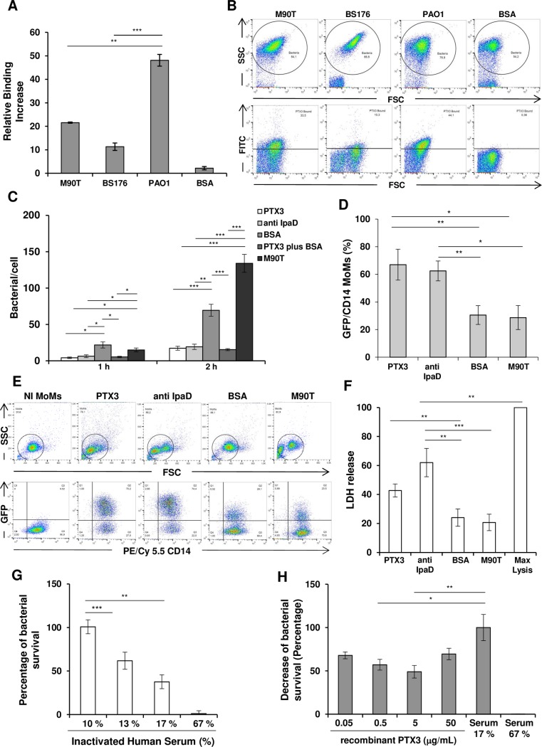
Fig 1. Opsonization of S. flexneri by PTX3 and downstream effects on cell populations and serum sensitivity.
(A) Binding of PTX3 to S. flexneri M90T and BS176 or P. aeruginosa (PAO1), was shown using biotin-labeled PTX3 (50 μg/mL, corresponding to 1,1 μM) or BSA (at 70 μg/mL), which was used as a control and evaluated by flow cytometry. (B) Representative flow cytometry data of three individual experiments of binding assay shown in panel A. PTX3-opzonized-bacteria display an increased FITC staining with respect to bacteria incubated with BSA. (C) Effect of PTX3 opsonization on HeLa cells. S. flexneri M90T was incubated for 1 h with either recombinant PTX3 (50 μg/mL, corresponding to 1,1 μM), anti-IpaD Ab, BSA (both at 50 μg/mL) or BSA plus recombinant PTX3 as above or left in medium alone (M90T) and used to infect HeLa cells at a multiplicity of infection (MOI) of 50. The number of bacteria per infected cell was evaluated at 1 h and 2 h of incubation post-infection (p.i.). (D, E and F) Effect of PTX3 opsonization on human macrophages (MoMs). (D) Bacterial internalization in MoMs. M90T (expressing GFP) treated with either recombinant PTX3 (50 μg/mL, corresponding to 1,1 μM), anti-IpaD Ab or BSA (both at 50 μg/mL) as above was used to infect MoMs at MOI 5 for 2 h p.i. M90T incubated in medium alone was used in parallel (M90T) (E) Representative flow cytometry data of three individual experiments of bacterial internalization in MoMs shown in panel D. The cells were stained for CD14+ (PE/Cy 5.5) and evaluated by flow cytometry. CD14+ GFP+ cells were counted as infected cells. Opzonized-bacteria display an increased CD14+ GFP+ staining with respect to untreated bacteria or bacteria treated with BSA. (F) Lactate dehydrogenase (LDH) release in supernatant of MoMs treated as above. (G) Serum bactericidal assay: 105 CFU/mL of exponential phase M90T were treated with varying concentrations of human serum (NHS) or heat-inactivated serum (HIS). % survival: CFU in the presence of NHS/CFU in the presence of HIS x 100. (H) Serum bactericidal assay as in (E) in the presence of PTX3. Bacteria were exposed to NHS (17%) in the presence of varying concentrations of PTX3 and survival was calculated as % of reduction of CFU number at 17% of NHS plus PTX3/ number of CFU at 17% NHS taken as value of 100. 0.5 μg/mL PTX3 vs 17% serum (no PTX3) p 0.05; 5 μg/mL PTX3 vs 17%serum (no PTX3) p 0.01. Data reported are the mean values (± SEM) of three independent experiments. (* p < 0.05; ** p < 0.01; *** p < 0.001 with Student’s t-test).

