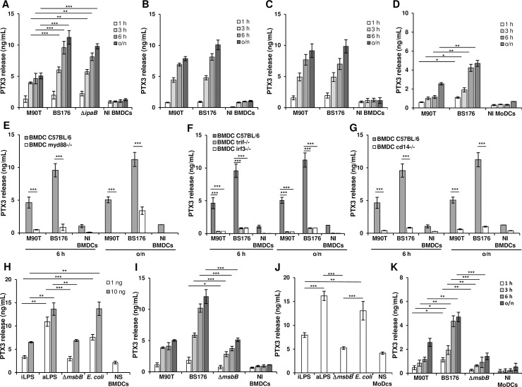
Fig 4. Production of PTX3 by S. flexneri in C57BL/6 BMDCs and MoDCs.
(A) PTX3 release in supernatant of infected BMDCs. Cells were infected with the invasive S. flexneri strain M90T or with the non-invasive BS176 or M90T ΔipaB at MOI 10 for 1 h, 3 h, 6 h and 18 h p.i. (o/n) (B) BMDCs infected as in (A) with M90T and BS176 previously treated with gentamicin (60 μg/mL for 1 h). (C) BMDCs treated with cytochalasin D (0.4 μg/mL for 1 h) and then infected with M90T and BS176 as above. (D) MoDCs infected with M90T and BS176 at MOI 10 for 1 h, 3 h, 6 h and (o/n) p.i. (E, F, G) BMDCs from (E) Myd88-/-; (F) Trif-/- and Irf3-/- and (G) Cd14-/- mice infected with M90T and BS176 as above. (H) BMDCs stimulated with 1 ng and 10 ng of LPS derived from intracellular shigellae (iLPS), shigellae grown in TSB medium (aLPS), Shigella ΔmsbB1 ΔmsbB2 (ΔmsbB) LPS and purified commercial E. coli LPS for 12 h. (I) BMDCs infected with M90T or BS176 or M90T ΔmsbB1 ΔmsbB2 at MOI 10 as in (A). (J) MoDCs stimulated with 1 ng and 10 ng of LPSs as in (H); (K) MoDCs infected with M90T or BS176 or M90T ΔmsbB1 ΔmsbB2 at MOI 10 as in (D). Graphs show the mean ± SD of triplicate wells and are representative of three independent experiments (* p < 0.05, ** p < 0.01, *** p < 0.001, Student’s t-test). NI: Not infected; NS: Not stimulated.

