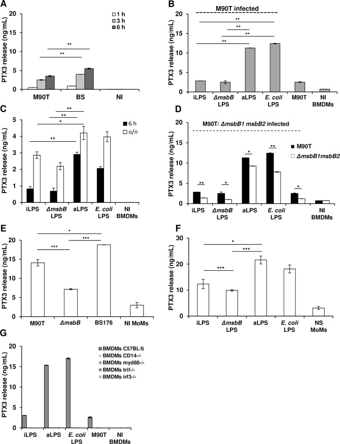
Fig 5. PTX3 production in S. flexneri infected C57BL/6 BMDMs and MoMs.
(A) PTX3 release in supernatant of BMDMs after infection with the S. flexneri strain M90T and its non-invasive derivative BS176 (MOI 10) at 1 h, 3 h and 6 h p.i. (B) BMDMs stimulated with 10 ng/mL of LPS derived from intracellular shigellae (iLPS), shigellae grown in TSB medium (aLPS), M90T ΔmsbB1 ΔmsbB2 LPS (ΔmsbB) and purified commercial E. coli LPS for 4 h, and infected with M90T (MOI 10) for 3 h p.i. (C) BMDMs after stimulation with 10 ng/mL of LPSs as in (B), at 6 h and 18 h (o/n). (D) BMDMs stimulated with 10 ng/mL of LPSs as in (B) for 4 h, and infected with M90T ΔmsbB1 ΔmsbB2 (MOI 10) for 3 h p.i. (E) MoMs infected with Shigella M90T, M90T ΔmsbB1 ΔmsbB2 and BS176 (MOI 0.1) for 3 h p.i. (F) MoMs after stimulation with 0.5 ng/mL of LPSs as in (B) for 12 h. (G) Myd88-/-, Trif-/-, Irf3-/-, Cd14-/- and wild type BMDMs pre-stimulated and infected with M90T as described in (B). Graphs show the mean ± SD of triplicate wells and are representative of three independent experiments (* p < 0.05, ** p < 0.01, *** p < 0.001). NI: Not infected; NS: Not stimulated.

