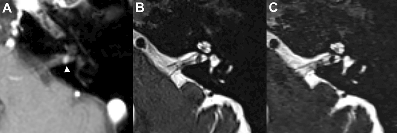
Figure 1–

Axial post-contrast T1-weighted MRI demonstrates a 3-mm vestibular schwannoma in the posterior left internal auditory canal (A, white arrowhead). All 3 radiologists blinded to the post-contrast images independently detected this mass on the isolated companion axial conventional (B) and compressed-sensing T2 SPACE images (C)(acquisition time = 250 & 50 seconds respectively). Side-by-side comparison demonstrates more noise in the compressed sensing SPACE within the temporal bone and cerebellar parenchyma, however there is adequate detail for screening assessment of the cerebellopontine angle, internal auditory canal and fluid-filled inner ear structures.
