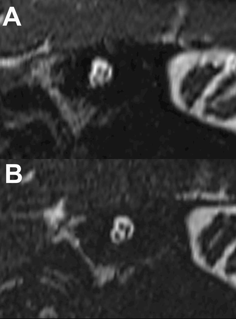
Figure 3–

Stenver’s view reconstructed image of the internal auditory canal using conventional and compressed-sensing T2 SPACE sequences (panels A & B respectively). There is less blurring at the bony margins of the IAC and less blurring of the cochlear, facial and vestibular nerves with the compressed sensing SPACE acquisition. Because this acquisition still takes 50 seconds, the motion degradation differences observed were more likely due to decreased macroscopic head motion and not related to CSF pulsatility in the internal auditory canal.
