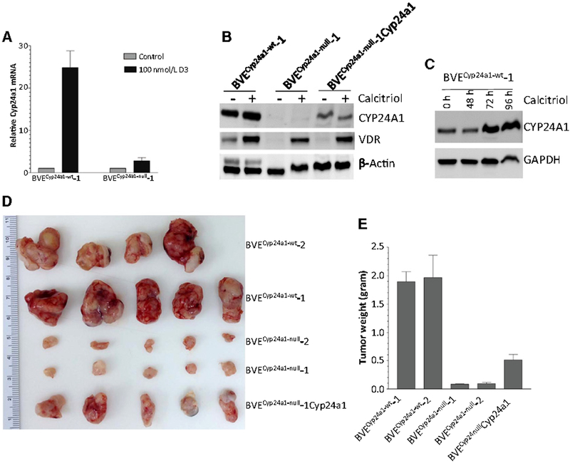
Figure 2.
Tumorigenicity of BVECyp24a1-null-derived tumor cells. A, Loss of Cyp24a1 expression in the BVECyp24a1-null-derived tumor cells. Cyp24a1 expression was analyzed by qRT-PCR in cells cultured in the presence or absence of 100 nmol/L calcitriol for 16 hours. The data are expressed as relative expression of Cyp24a1 after normalization to β-actin expression in each sample. Results of controls were adjusted as 1. B, Western blot analysis of CYP24A1 and vitamin D receptor (VDR) expression in BVECyp24a1-wt-1, BVECyp24a1-null-1, and BVECyp24a1-null-1 cells transfected with wild-type Cyp24a1 (BVECyp24a1-null-1Cyp24a1). The cells were cultured in the presence or absence of 100 nmol/L calcitriol for 72 hours. C, Time course of CYP24A1 protein expression after calcitriol stimulation. BVECyp24a1-null-1 cells were cultured in the presence of 100 nmol/L calcitriol for 48, 72, and 96 hours, respectively. The CYP24A1 protein expression was detected by Western blot analysis. D, Tumor growth in nude mice following subcutaneous injection of 2 × 10⁶ tumor cells from each of BVECyp24a1-wt-1, BVECyp24a1-wt-2, BVECyp24a1-null-1, BVECyp24a1-null-2, or BVECyp24a1-null-1Cyp24a1. Four weeks after injection, the tumors were removed, and their weights were measured. E, Tumor weight in each group at 4 weeks after injection. Tumorigenic potential was significantly reduced after the loss of CYP24A1 expression. The tumor growth potential was partially restored in the BVECyp24a1-null-1 cells following transfection of wild-type Cyp24a1 expression plasmid.

