Fig. 1.
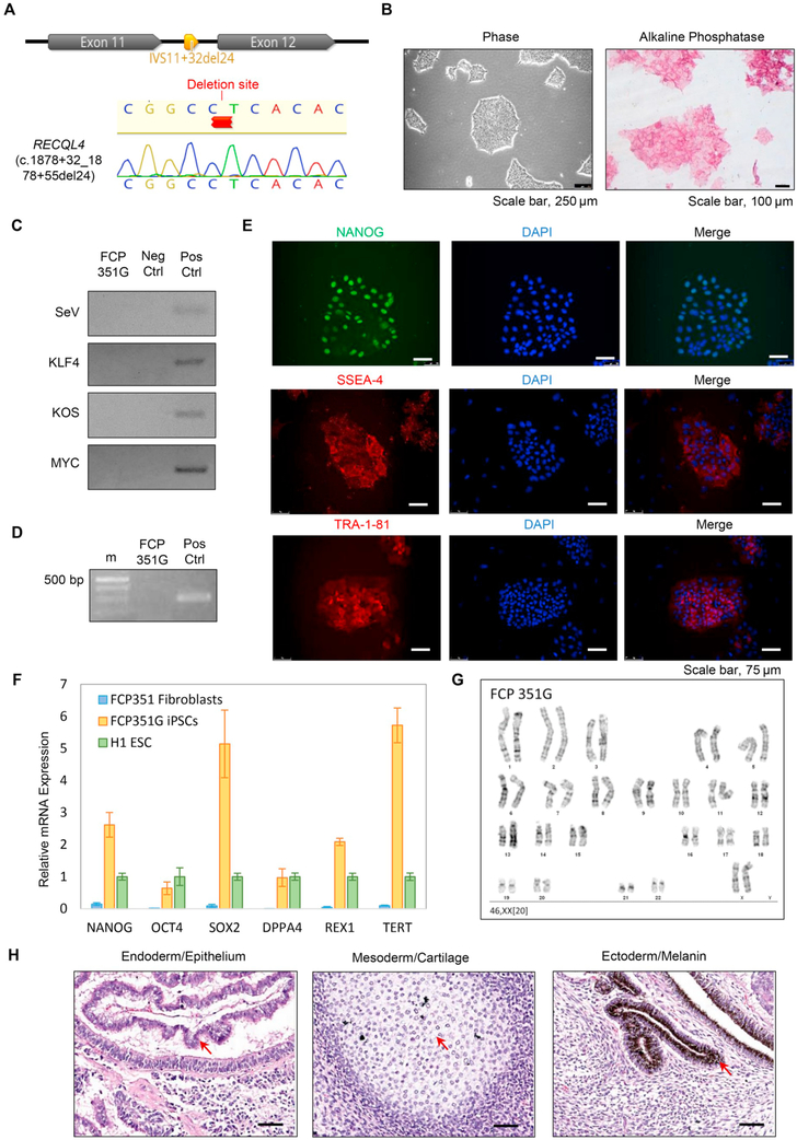
Generation and characterization of FCP351G iPSCs. (A) Sanger sequence of RECQL4 Exons 11-12 depicting the deletion found in FCP351 fibroblasts and verified by Sanger sequencing (see chromatogram) in FCP351G iPSCs. (B) Cell morphology (left panel) and alkaline phosphatase staining (right panel) of the FCP351G line. Scale bar, 250 μm (left) and 100 μm (right). (C) PCR detection of exogenous OCT4, SOX2, KLF4 and c-MYC in FCP351G iPSCs. (D) Mycoplasma PCR assay in FCP351G iPSCs.(E) Immunofluorescence staining of pluripotency factor NANOG and hESC surface markers (SSEA4 and TRA-1-81) in the FCP351G line. Scale bar, 75 μm. (F) qRT-PCR assay for the expression of endogenous pluripotency genes (NANOG, SOX2, OCT4, DPPA4, REX1, and TERT) in the FCP351G iPSCs. (G) A representative G-banded karyotype from iPSCs (FCP351G) showing 46, XX. (H) In vivo teratoma assay of the FCP351G iPSCs. Scale bar, 50 μm.

