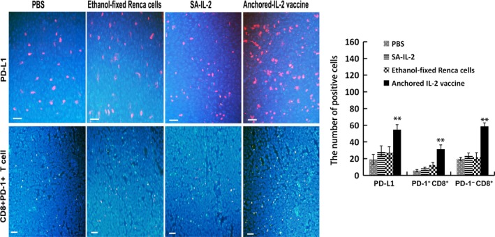
Figure 2.

Programmed death receptor‐1 (PD‐1)/programmed death ligand‐1 (PD‐L1) signaling exists in the tumor microenvironment after interleukin (IL)‐2‐modified vaccine therapy. Immunofluorescence assays were carried out to evaluate PD‐L1 expression and infiltration of PD‐1+ CD8+ T cells in the tumor tissues. DAPI (blue fluorescence) stains the cell nucleus, and tumor cells are presented as blue fluorescence. Red fluorescence represents PD‐L1 positive cells, green fluorescence represents CD8+ T cells, and yellow fluorescence represents PD‐1+ CD8+ T cells (Bar, 200 μm). Results of statistical analyses showed significant differences in the number of positive cells (n = 15). All the experiments were replicated at least three times (**P < .01)
