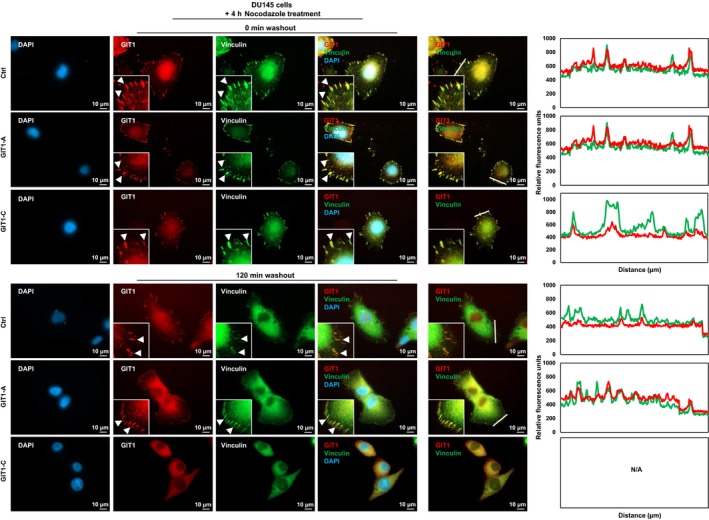
Figure 5.

Differential functions of the GIT1 splice variants in FA stability. DU145 stable cell lines overexpressing GIT1‐A, GIT1‐C, or empty vector were seeded on coverslips and serum‐starved. They were treated with 10 μmol/L nocodazole for 4 h, subsequently washed away, and replaced with serum‐containing medium. Cells were fixed at 0 or 120 min after the washout, costained against GIT1 and vinculin, and then mounted with DAPI staining mount. Cells were imaged using a Zeiss AxioObserver Z1 (Carl Zeiss AG; Oberkochen, Germany) microscope, where the scale bar represents 10 μm. Arrowheads indicate FA complexes. Overlapping signals between GIT1 and vinculin appear yellow. Overlapping of the two signals in a cross‐section (indicated by white line) of FA complexes were profiled by the ZEN program. All experiments were repeated three times. FA, focal adhesion; GIT1, G‐protein‐coupled receptor kinase‐interacting protein 1; IF, immunofluorescence; ZEN, ZEISS efficient navigation
