
Figure 1.
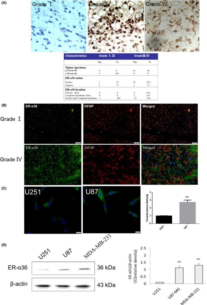
ER‐α36 was overexpressed in glioblastoma specimen. A, Immunohistochemistry stained ER‐α36 expression in human glioblastoma. B, Immunofluorescence (IF) staining of ER‐α36 (green) and anti‐glial fibrillary acidic protein (GFAP) (red) in human glioblastoma. Nuclei were counterstained with DAPI (blue). C, IF staining of ER‐α36 in U87 and U251 cells (green). Nuclei were counterstained with DAPI (blue). D, Western blot analysis shows the expression of ER‐α36 in U87 and U251 cells, with β‐actin as internal control. (n=3‐5, **P < 0.01) ER, estrogen receptor