
Figure 5.
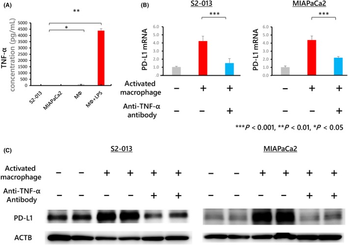
A, The concentration of tumor necrosis factor (TNF)‐α in each conditioned medium determined by ELISA. Macrophages treated with lipopolysaccharide (LPS) produced substantial amounts of TNF‐α. B, Human monocyte‐derived macrophages were co‐cultured with S2‐013 or MIAPaCa2 cells. An anti‐TNF‐α antibody was then used at 1 μg/mL to neutralize TNF‐α produced by macrophages treated with 100 ng/mL LPS for 24 h. PD‐L1 mRNA expression in S2‐013 and MIAPaCa2 cells was upregulated after co‐culture with activated macrophages; this upregulation was inhibited by treatment with 1 μg/mL anti‐TNF‐α antibody. The plus symbol indicates that the antibody was added, and the minus symbol indicates that the antibody was not added. C, Western blot analysis of proteins detected by probing with anti‐PD‐L1 antibodies. PD‐L1 expression upregulated by co‐culture with activated macrophages was inhibited by an anti‐TNF‐α antibody. Full‐length gels are presented in Figure S5