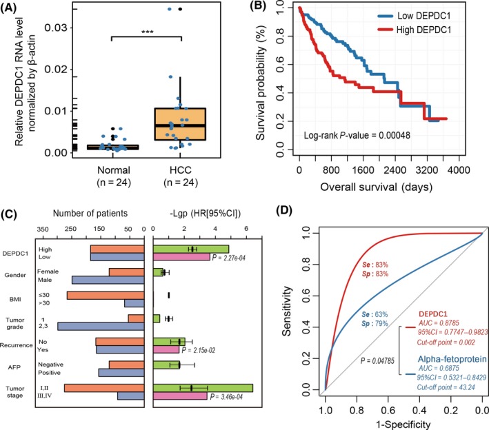
Figure 2.

Differential DEP domain containing 1 (DEPDC1) expression level shows diagnostic and prognostic significance in liver hepatocellular carcinoma (LIHC) samples. A, Expression level of DEPDC1 in 24 paired hepatocellular carcinoma (HCC) tissues. B, Overall survival of The Cancer Genome Atlas (TCGA) LIHC patients with DEPDC1 expression level. P‐values were calculated using the log‐rank test. C, Risk evaluation of DEPDC1 and clinical parameters. Left bar panel shows the number of patients in different groups divided by the status of each clinical parameter. Right bar panel shows the P‐value (−log10 transformed) and hazard ratio of univariate Cox regression for each clinical parameter. The clinical parameter with a significant P‐value (< .01) was selected to construct multivariate Cox regression, and the significant P‐value is indicated beside the corresponding bars (pink). BMI, body mass index. D, Receiver operating characteristic curve of DEPDC1 expression level contrasted with alpha‐fetoprotein (AFP; ng/mL) on 24 paired HCC tumor and normal liver tissues. ***P < .001
