Figure 1.

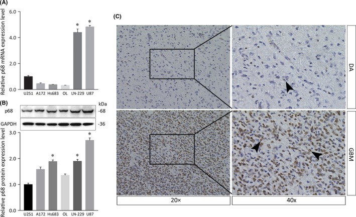
Expression of p68 in various glioma cells and immunolocalization of p68 in diffuse astrocytoma and glioblastoma. A, Relative mRNA expression levels of the p68 gene (p68 mRNA:GAPDH mRNA ratios) in U251, A172, Hs683, OL, LN‐229, and U87 glioma cell lines were calculated by quantitative real‐time PCR. B, Western blot analysis showing the p68 protein in glioma cell lines. Histogram of relative p68 protein expression with relative ratios shown as percentages of the p68/GAPDH band. Bars represent SD; *P < .05. C, Immunolocalization of p68 in diffuse astrocytoma (DA; upper panels) and glioblastoma (GBM; lower panels) tissues. Note that p68 immunolocalizes to neoplastic astrocytes (right panels, arrowheads). Strong staining was detected in GBM tissues (lower panels), whereas faint staining was observed in DA tissues (upper panels)
