
Figure 4.
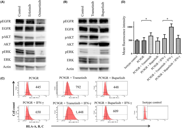
Effects of MEK and PI3K inhibitors on major histocompatibility complex class I (MHC‐I) expression in PC9GR cells. A,B, Cells were incubated for 6 h in the absence or presence of erlotinib (50 nmol/L) or osimertinib (50 nmol/L) (A) or of trametinib (100 nmol/L) or buparlisib (500 nmol/L) (B), after which cell lysates were subjected to immunoblot analysis with antibodies to phosphorylated (p) or total forms of epidermal growth factor receptor, AKT, or extracellular signal‐regulated kinase or with those to β‐actin (loading control). C,D, Cells were incubated with or without trametinib (100 nmol/L) or buparlisib (500 nmol/L) and in the absence or presence of interferon‐γ (20 U/mL) for 48 h, after which cell surface expression of MHC‐I was analyzed by flow cytometry with antibodies to a shared epitope of HLA‐A, ‐B, and ‐C. Representative flow cytometric profiles (C) and the mean fluorescence intensity (MFI) of MHC‐I–positive cells determined as means + SE from three independent experiments (D) are shown. *P < 0.05 (unpaired Student's t test)