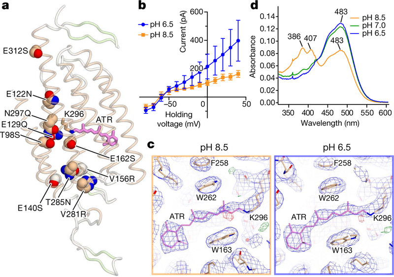
Fig. 1 |. Structures of iC++ and insights into pH dependence.
a, Crystal structure of iC++ at pH 8.5. iC++ mutations are shown with sphere models. b, pH-dependent photocurrents of iC ++ at pHext 6.5 (blue) and 8.5 (orange). Data are mean ±s.e.m. of 5 cells. c, Retinal-binding pocket (RBP) of iC++ at pH 8.5 and 6.5; 2Fo − Fc maps (blue mesh, contoured at 1σ) and Fo − Fc maps (green and red meshes, contoured at 3σ and −3σ, respectively) are shown. d, iC++ absorbance spectra at pH 6.5, 7.0 and 8.5.

