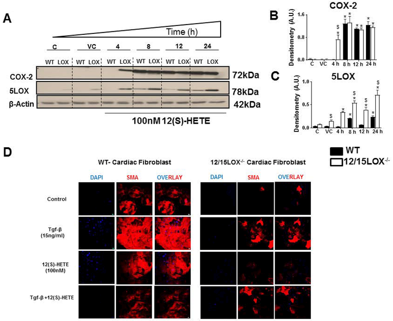
Figure 3: 12(S)-HETE promotes early induction of COX-2 and 5LOX in cardiac fibroblast and trans-differentiation of fibroblast to myofibroblast.
Cardiac fibroblast treated with 12(S)-HETE (100nM) for 4, 8, 12 and 24 h. A Immunoblot representing COX-2 and 5LOX expression in WT and 12/15LOX−/− cardiac fibroblast treated with in 12(S)-HETE. B Densitometric analysis of COX-2 levels. C Densitometric analysis of 5LOX levels. *p<0.05 vs untreated cells, $p<0.05 vs. treated WT cells. Results are presented as mean ± SEM. Data are representative of a n=3 independent experiments. D Representative immunofluorescence images representing α-SMA expression (Red) and Hoechst (blue) in cardiac fibroblast isolated from WT and 12/15LOX−/− with and without treatment of Tgf-β (15ng/ml) and 12(S)-HETE (100nM) for 18 h. Data are representative of n=3 independent experiments.

